A new machine that helps scientists speedily analyse single cells from individual people could bring hope to millions of children suffering from rare blood disorders such as sickle cell disease.
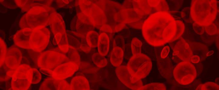
Red blood cells are used to carry oxygen around the body and damage to them can lead to life-threatening conditions. Image: Shutterstock/Fenton
It is part of a number of new technologies that are helping to diagnose, treat and even prevent lethal anaemias, disorders in which people’s red blood cells are unable to efficiently transport oxygen around the body.
Over 300 000 babies are born each year with sickle cell disease, a genetic defect that causes red blood cells to bend, stick and plug up narrow blood vessels. This prevents enough oxygen from being carried in the blood stream, leading to acute pain that can last for days and life-threatening oxygen deficiency in vital organs.
“
‘There are still forms of anaemia for which we have no clue as to their cause.’
In high-income countries, only a handful of costly treatments can extend the life expectancy of patients. In sub-Saharan Africa, where the mutation is widespread, children with sickle-cell disease tend to die before their fifth birthday.
The EU-funded CoMMiTMenT project brings together academics, companies and medical centres to build a machine that can rapidly spot and analyse sick blood cells and help scientists tailor personalised treatments for them.
Dr Lars Kaestner from Saarland University in Germany explains that diagnosing and treating rare anaemias is challenging because of low patient numbers and high drug development costs.
‘Some of these diseases are so rare that scientists would need to study the cells of each patient individually to develop treatments for them,’ he said. ‘This is just not affordable with current technology.’ Even conditions that afflict millions of people do not always find a compelling business case.
Dr Kaestner, who coordinates the CoMMiTMenT project, says the new machine, called µCOSMOS, combines different molecular imaging technologies developed by a number of European SMEs in order to inspect hundreds of millions of cells an hour and automate the diagnosis process.
µCOSMOS performs an initial screening process with optical microscopes and image recognition software to identify red blood cells with irregular shapes or sizes in real time. An infrared laser then prods these cells under a flurorescence microscope to inspect properties such as their surface composition and how they interact with other cells.
Cells that display potentially harmful traits are studied in depth using scanning ion conductance microscopy. ‘Scanning ion conductance microscopy can reveal the surface structure and membrane properties of each cell,’ said Dr Kaersten. ‘It sweeps their surface with a microscopic electrode that measures nanoscopic nuances in conductivity.’
Dr Anna Bogdanova, a physiologist at the University of Zurich, Switzerland, is helping to combine these technologies into an instrument that can be used by medical practitioners.
Microfluidic chips
Microfluidic chips are tiny obstacle courses carved out of plastic, which guide fluids using differences in the force of adherence to their sidewalls.
According to Professor Charles Baroud at the Ecole Polytechnique in Paris, France, microfluidic chips offer an advantage over conventional testing platforms in that they can fix statistically representative batches of cells into place so that researchers can repeat measurements on them indefinitely.
In separate work funded by the EU’s European Research Council, Professor Baroud is developing microfluidic chips that break liquids down into tens of thousands of nanolitre droplets, and align them onto areas half the size of a credit card.
He has already shown how sickled red blood cells degenerate over time and expects further work to help unlock the secrets of the disease. He is now working with biologists on a range of cellular disorders and pathogens.
Dr Kaestner says the ability to analyse the state of single cells is an important development in diagnosing anaemias. ‘There are still forms of anaemia for which we have no clue as to their cause,’ he said. ‘This growing ability to study single cells in single patients offers new hope in treating rare and overlooked diseases.’
A universal cure?
Dr Marina Cavazzana, at INSERM in France, is addressing the challenge of hereditary anaemias from a different angle. Rather than finding ways to counteract the damage caused by genetic mutations, her team aims to uproot the mutations themselves, so that red blood cells are born healthy.
This means editing the DNA of the stem cells in the bone marrow of patients. Her team and partners, which include the biotech firm Bluebird, have engineered a benign virus to enter the nucleus of these cells and recode faults in their DNA with a healthy genetic sequence.
Gene therapy is not yet a low-cost option and the technique is still undergoing clinical trials, but unlike other treatments explored to date, it offers the prospect of a universal cure for sickle cell disease and other genetic disorders.
A microscope developed by the CoMMiTMenT project screens millions of red blood cells and isolates anomalies for further testing by prodding them out of the viewing plane with an infrared laser. Video courtesy: Dr Lars Kaestner

